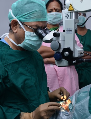
Our Centre is equipped with a State-of-the-art Operation Theatre for a wide range of Ophthalmic Surgery. A surgery is usually an in-patient procedure where the patient needs to remain in observation for only a couple of hours after the surgery is completed.
We perform the following Surgeries in our facility using the Latest Technology.
What is a Cataract?
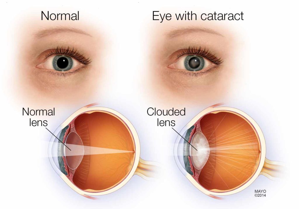
A cataract is an eye disease that affects a large percentage of population. It is a clouding of the natural crystalline lens in the eye, and is made of water and protein. Usually, by age 65, the chemical changes in our body cause the lens to grow cloudy or yellow, preventing light rays from focusing properly inside our eye. When untreated, it can cause vision loss. 80% of curable blindness is due to cataract.
How to recognize Cataract?
Does your vision seem cloudy or hazy to you?
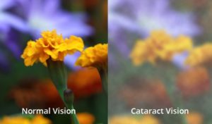
The cataract starts out small and can slowly start affecting your vision. The vision gets blurred or cloudy, and the eyes may get sensitive to bright light.
See your doctor right away before the condition worsens. Need an appointment.. Contact us today.
Micro Incision Cataract Surgery
A cataract can worsen if not removed. Surgery is the only option to remove it. And we can remove the cataract whenever it begins to affect your vision and thereby your lifestyle.
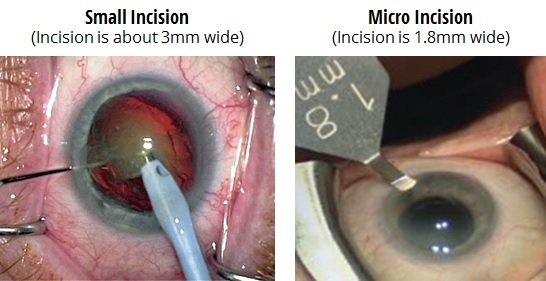
At Jaya Eye Care, our Chief Doctor uses an advanced technique called Microincision Cataract Surgery. This procedure provides greater control and precision and the post operative outcomes are much better than standard procedures (Small Incision Cataract Surgery where the incision is above 3mm wide).
Reducing the incision width to 1.8mm, which is also called Microincision, means less invasiveness, faster healing and quicker recovery. The patient can return back to normal life quickly.
Cataract Surgery Procedure at Jaya Eye Care
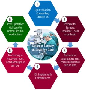
We will first evaluate your eyes and provide Counselling to preparing you for surgery and choosing a new lens also called Intraocular Lens (IOL) for implant.
An IOL is a clear, plastic lens that requires no care and becomes a permanent part of your eye. Light is focused clearly by the IOL onto the retina, improving your vision. You will not feel or see the new lens. You can select from a wide choice of IOLs based on physiological and occupational needs.
The surgery is an in-patient procedure and you will be given local anaesthesia. Our Eye Surgeon will first remove the cataractous lens using a procedure called Phacoemulsification. It will be replaced with a Foldable IOL in your eye. This is a permanent implant and the lens does not degrade.
The whole procedure usually takes 30 minutes. You will be monitored in our recovery room for a couple of hours and then you will be discharged right away. With proper post operative care, you can get back to normal life in a week’s time.
We also perform Small Incision Cataract Surgery in certain cases.
Need Counselling for Surgery? Contact us. We will be happy to guide you.
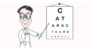
Cataract - Care and Act
Here is an interesting article: Cataract – Care and Act.
Have health insurance?
Feel free to contact our Insurace desk. Here is an article on Understanding Medical Insurance.
What is Squint?
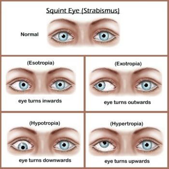
Squint (Strabismus) happens when the two eyes fail to maintain proper alignment and work together as a team. Though it is a common condition among younger population, affecting 2 to 4 percent of children, it may also appear later in life. The misalignment may be permanent or it may be temporary, occurring occasionally. The deviation may be in any direction: inward, outward, upward or downward.
How to recognize Squint?
The first sign of squint is a visible misalignment of the eyes, with one eye turning in, out, up, down or at an oblique angle.
See your doctor right away. In some cases they can be corrected with prescription glasses, other cases may need surgery. Contact us today for an appointment..
Surgical treatment of Squint (Paediatric and Adult)
Our doctors can treat squint non-surgically by Prescription Glasses, if they are caused by refractive errors.
Strabismus (Squint) surgery is very effective in most cases of squint. Treating squint early on with Surgery helps the affected eye in developing normal visual acuity and the two eyes functioning together as a team. Surgery is done under general anaesthesia in children and under local anaesthesia in cooperating adults.
What are Cornea related problems?
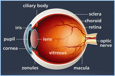
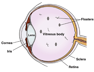
The cornea is your eye’s clear, outer layer at the front and center of the eye. It is transparent and permits light to pass into the eye, through the pupil, lens, and onto the retina at the back of the eye. Along with the sclera (the white of your eye), it serves as a barrier against dirt, germs, and other things that can cause damage.
If your cornea is damaged by disease, infection, or an injury, the resulting scars can affect your vision. They might block or distort light as it enters your eye.
Common corneal problems are corneal ulcers, keratitis (inflammation) and keratoconus (thinning), infections, allergies and corneal abrasions caused by injury.
How to recognize Cornea related problems?
You may notice symptoms like blurred vision, redness, extreme sensitivity to light. These symptoms may also mean other eye problems, that may be serious and requiring special treatment. Seek medical attention right away.
Need an appointment.. Contact us today.
Treatment options for Corneal related problems
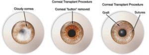
Treatment options include Corneal Transplant Surgery, Lamellar Corneal Surgery and Stem Cell Transplant and therapy, where the diseased cornea is removed and replaced with a natural cornea procured from a donor eye. Artificial cornea is also available now, for cases where multiple corneal transplants have failed.
Other than corneal surgery for corneal diseases, Refractive surgery for reduction of refractive error (glass number) and phakic intraocular lens implant surgery to correct myopia can be done.
What are Vitreoretinal Disorders?
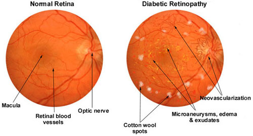
Vitreoretinal Disorders are eye conditions that involve the retina, macula, and vitreous humor (fluid) that fills the back of the eye. As these are at the back of the eye, they are not readily visible. Some of the conditions are diabetic retinopathy, macular degeneration, retinal detachments or tears, macular holes, retinopathy of prematurity, retinoblastoma, uveitis, eye cancer, flashes and floaters and retinitis pigmentosa.
These can affect people of any age or diabetics.
How to recognize Vitreoretinal Disorders?
Some of the visual symptoms of varying severity are floaters, which may be anything from small dots to large clouds of “debris”, a haziness of the entire visual field, and profound visual loss. See your eye doctor right away.
Contact us today for an appointment..
Treatment options for Vitreoretinal Disorders
Treatment by Laser procedures
At Jaya Eye Care Centre, we offer treatment by Laser procedures for Vitreoretinal Disorders.
Diabetic Retinopathy
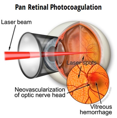
The retina may swell up caused by the damaged small vessels of the retina leaking fluid and blood. These changes decrease vision if the central part is affected. Laser is done to seal these leaks. However laser is done to prevent or retard further loss of vision and not to improve vision. In severe changes when new blood vessels have grown (Proliferative Diabetic Retinopathy), multiple sittings of laser are needed to regress these vessels. This is called Pan Retinal Photocoagulation and is highly effective in preventing severe visual loss due to recurrent bleeding in vitreous.
Retinal tears and holes
In a retinal tear or a hole without a retinal detachment, laser is done prophylactically to seal the hole and prevent or limit the detachment.
Age Related Macular Degeneration (AMD)
In patients with wet type of AMD not involving the central part, Laser can be done to close the Choroidal Neovascular Membrane. This can prevent severe visual loss. However in patients with central involvement, other specific lasers are used like- Photo Dynamic Therapy (PDT) with verteporfin and Transpupillary Thermo Therapy (TTT) with Diode laser.
Other Retinal vascular disorders
Retinal vein occlusions and Eales disease also require laser at times. In venous occlusions, the central retina may swell up due to fluid collection leading to reduced vision. Such cases require laser to decrease the swelling, which may help in improving the vision. Venous occlusions may also lead to new vessel formation, which needs laser to regress these vessels. Similarly in Eales disease, laser is required to reduce recurrent vitreous haemorrhages. Laser is also useful in selected cases with Central Serous Retinopathy (CSR).
Treatment by Surgery
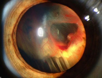
Retinal Detachment
Some of the causes of a detached retina are very high levels of myopia, injury to the eye or face. Our retinal specialists at Jaya Eye Care treat advanced cases like retinal detachments and vitreal hemorrhage by surgery.
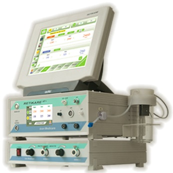
Vitrectomy is done to treat retinal detachment, where the vitreous gel is removed and usually combined with filling the eye with either a gas bubble or clear silicone oil. We perform Vitrectomy using advanced equipment called RETIKARE.
Equipment used in Vitreoretinal Surgery:
Iridex Diode Laser Model TX
Retikare Plus
Reti Cryo
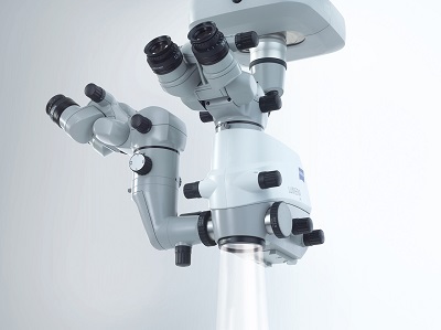
Carl Zeiss Lumera 300
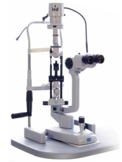
Slit Lamp Model SL 9900 3x LED

