What are Vitreoretinal Disorders?
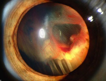
Vitreoretinal Disorders are eye conditions that involve the retina, macula, and vitreous humor (fluid) that fills the back of the eye also called the Posterior Segment of the eye. As these are at the back of the eye, they are not readily visible.
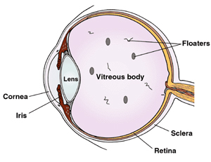
Some of the Vitreoretinal disorders are Diabetic Retinopathy, Macular Degeneration, Retinal detachments or tears, Macular holes, Retinopathy of Prematurity, Retinoblastoma, Uveitis, Eye cancer, Flashes and Floaters and Retinitis Pigmentosa.
These can affect people of any age or diabetics. Some of the conditions above are Diabetic Related Eye Disorders.
How to recognize Vitreoretinal Disorders?
Some of the visual symptoms of varying severity are floaters, which may be anything from small dots to large clouds of “debris”, a haziness of the entire visual field, and profound visual loss.
If you experience any of the above symptoms, see your eye doctor right away. Contact us today for an appointment.

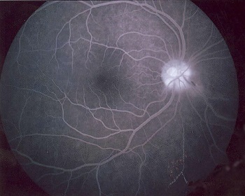
Evaluation
Treatment Options
Treatment by Laser procedures
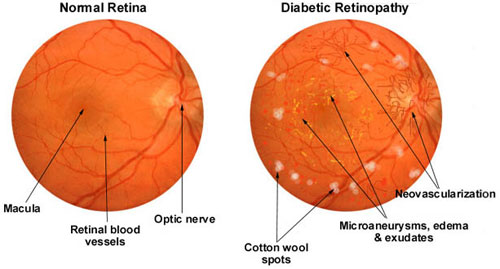
Diabetic Retinopathy
The retina may swell up caused by the damaged small vessels of the retina leaking fluid and blood. These changes decrease vision if the central part is affected.
Treatments for Diabetic retinopathy include replacement of the inner gel inside the eye and different kinds of laser surgery. Laser is done to seal these leaks. However laser is done to prevent or retard further loss of vision and not to improve vision.
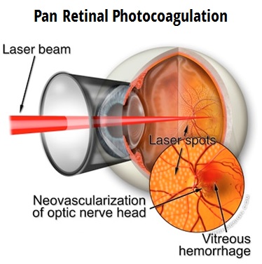
In severe changes when new blood vessels have grown (Proliferative Diabetic Retinopathy), multiple sittings of laser are needed to regress these vessels. This is called Pan Retinal Photocoagulation and is highly effective in preventing severe visual loss due to recurrent bleeding in vitreous.
Better control of blood sugar levels can slow down the progression of the disease.
Retinal tears and holes
In a retinal tear or a hole without a retinal detachment, laser is done prophylactically to seal the hole and prevent or limit the detachment.
Age Related Macular Degeneration (AMD)
In patients with wet type of AMD not involving the central part, Laser can be done to close the Choroidal Neovascular Membrane. This can prevent severe visual loss. However in patients with central involvement, other specific lasers are used like- Photo Dynamic Therapy (PDT) with verteporfin and Transpupillary Thermo Therapy (TTT) with Diode laser.
Other Retinal vascular disorders
Retinal vein occlusions and Eales disease also require laser at times. In venous occlusions, the central retina may swell up due to fluid collection leading to reduced vision. Such cases require laser to decrease the swelling, which may help in improving the vision. Venous occlusions may also lead to new vessel formation, which needs laser to regress these vessels. Similarly in Eales disease, laser is required to reduce recurrent vitreous haemorrhages. Laser is also useful in selected cases with Central Serous Retinopathy (CSR).
Treatment by Surgery
Retinal Detachment
Some of the causes of a detached retina are very high levels of myopia, injury to the eye or face. Our retinal specialists at Jaya Eye Care treat advanced cases like retinal detachments and vitreal hemorrhage by surgery.
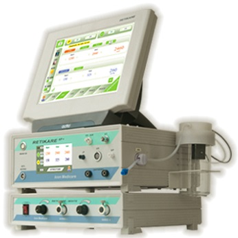
Vitrectomy is done to treat retinal detachment, where the vitreous gel is removed and usually combined with filling the eye with either a gas bubble or clear silicone oil. We perform Vitrectomy using advanced surgical equipment called RETIKARE.
Equipment used in Vitreoretinal Surgery:
- Iridex Diode Laser Model TX
- Retikare Plus
- Reti Cryo
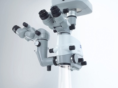
- Carl Zeiss Lumera 300
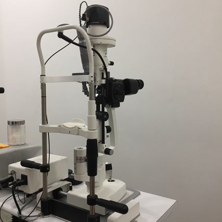
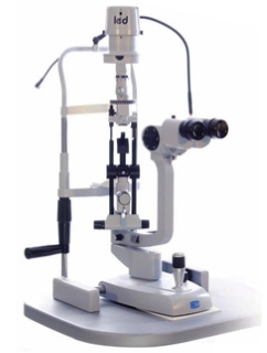
- Slit Lamp Model SL 9900 3x LED
Experiencing blurred, reduced or cloudy vision or eye strain, redness or itching?
Seek medical attention now. Get your eyes evaluated and treated. Need an appointment.. Contact us today. Get your Eye Health back on track!

