Our Eye Specialists and Optometrists are highly skilled and experienced to do thorough Eye Investigations at Jaya Eye Care. We can diagnose and monitor Eye diseases and conditions with the aid of our latest Equipment.
Following are the Eye Investigations we conduct at our centre.
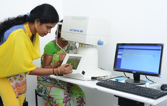
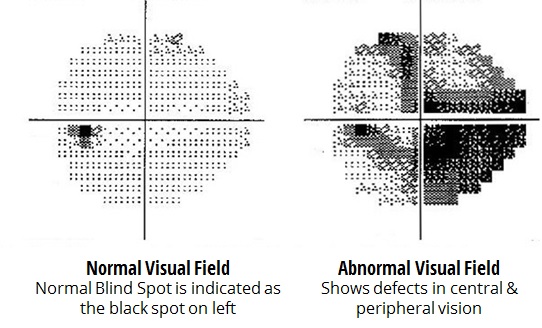
The patient is made to sit and look inside the perimeter equipment and asked to press a button each time they see a flash of light. A computer records the spot of each flash and also if the patient pressed the button when the light flashed in that spot. Based on this reading, a report is generated on the areas where the eye did not perceive the light flash, indicating areas of vision loss. This usually indicates an early sign of glaucoma.
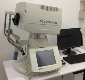
We use the latest Equipment, called Perimeter Model Octopus 300 with IT 2000, which is completely automated. This aids diagnostics by allowing a detailed printout of the patient’s visual field.
Equipment used for Visual Fields Test:
Perimeter Model Octopus 300 with IT 2000
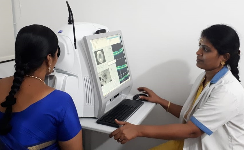
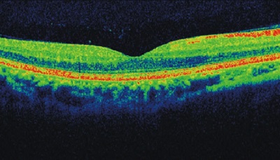
Optical coherence tomography (OCT) is a non-invasive imaging test that uses light waves to take cross-section pictures of your retina, the light-sensitive tissue lining the back of the eye.
With OCT, each of the retina’s distinctive layers can be seen, allowing your ophthalmologist to map and measure their thickness. These measurements help with diagnosis and provide treatment guidance for glaucoma and retinal diseases, such as age-related macular degeneration and diabetic eye disease.
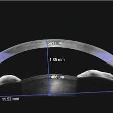
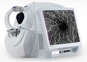
Our CIRRUS HD-OCT Model 500 by Carl Zeiss Vision has clinical assessment tools that offers imaging applications for anterior segment, glaucoma and retina.
The Anterior Segment Imaging module includes a wide view of the entire anterior chamber.
Early glaucoma, diabetic retinopathy and macular changes can be diagnosed with the high definition OCT scans using multiple modules.
Equipment used for Optical Coherence Tomography (OCT):
CIRRUS HD-OCT Model 500 by Carl Zeiss Vision
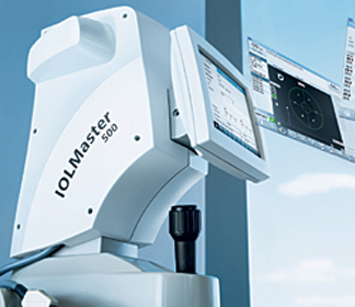
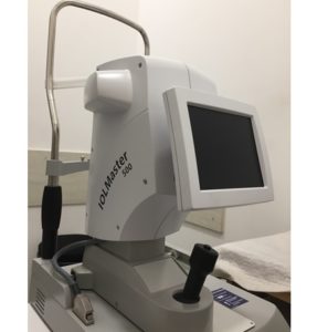
The IOL Master allows for a non contact, non invasive method for determining the power of the Intraocular lens (IOL) to be implanted for Cataract Surgery.
The IOL Master provides fast, accurate measurements of eye length and surface curvature, necessary for Mono-focal, Multi-focal, Toric IOLs in cataract surgery. The IOL Master is very efficient and accurate. Also, because the IOL Master is non-contact (nothing touches the eye itself), there is no need for anesthesia and there is no potential for spread of contamination from the IOLMaster.
This Optical Biometer helps with advanced measurement of challenging eyes with denser cataracts.
Equipment used for measurement of IOL power:
Carl Zeiss IOL Master 500
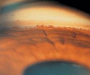
Gonioscopy is an eye investigation to look at the front part of your eye (anterior chamber) between the cornea and the iris. Gonioscopy is a painless examination to see whether the area where fluid drains out of your eye is open or closed.
It is done during regular eye evaluation, depending on the patient age, especially if they at high risk for glaucoma.
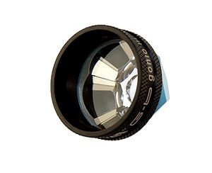
A goniolens or gonioscope is used in conjunction with a slit lamp or operating microscope to view the anatomical angle formed between the eye’s cornea and iris. It helps in diagnosing and monitoring various eye conditions associated with glaucoma.

